Introduction
The distribution of the cardiac output to various parts of the body at rest in a normal man. The general principles described in the preceding chapters apply to the circulation of all these regions, but the vascular supplies of many organs have additional special features that are important to their physiology
Cerebral circulation: anatomic considerations
The principal arterial inflow to the brain in humans is via four arteries: two internal carotids and two vertebrae. In humans, the carotid arteries are quantitatively the most significant. The vertebral arteries unite to form the basilar artery, and the basilar artery and the carotids from the circle of Willis below the hypothalamus. The circle of Willis is the origin of the six large vessels supplying the cerebral cortex.
Read More: themakernewsz.com
Formation & absorption
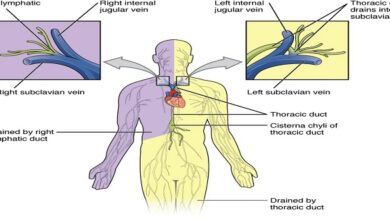
CSF fills the ventricles and subarachnoid space. In humans, the volume of CSF is about 150 mL and the rate of CSF production is about 550 mL/d. Thus the CSF turns over about 3.7 times a day. In experiments on animals, it has been estimated that 50– 70% of the CSF is formed in the choroid plexuses and the remainder is formed around blood vessels and along ventricular walls. Presumably, the situation in humans is similar. The CSF in the ventricles flows through the foramina of Magnesia and Lustra to the subarachnoid space and is absorbed through the arachnoid villi into veins, primarily the cerebral venous sinuses. The villi consist of projections of the fused arachnoid membrane and endothelium of the sinuses into the venous sinuses. Similar, smaller villi project into veins around spinal nerve routes. These projections may contribute to the outflow of CSF into venous blood by a process known as bulk flow, which is unidirectional.
Protective function
The most critical role for CSF (and the meninges) is to protect the brain. The Dura is attached firmly to the bone. Normally, there is no “subdural space,” with the arachnoid being held to the Dura by the surface tension of the thin layer of fluid between the two membranes. The brain itself is supported within the arachnoid by the blood vessels and nerve roots and by the multiple fine fibrous arachnoids trabecular. The brain weighs about 1400 g in air, but in its “water bath” of CSF, it has a net weight of only 50 g. The buoyancy of the brain in the CSF permits its relatively flimsy attachments to suspend it very effectively. When the head receives a blow, the arachnoid slides on the Dura, and the brain moves, but its motion is gently checked by the CSF cushion and by the arachnoid trabecular.
Head injuries
Without the protection of the spinal fluid and the meninges, the brain would probably be unable to withstand even the minor traumas of everyday living; but with the protection afforded, it takes a fairly severe blow to produce cerebral damage. The brain is damaged most commonly when the skull is fractured and bone is driven into neural tissue (depressed skull fracture), when the brain moves far enough to tear the delicate bridging veins from the cortex to the bone, or when the brain is accelerated by a blow on the head and is driven against the skull or the tentorium at a point opposite where the blow was struck.
Circumventricular organs
When dyes that bind to proteins in the plasma are injected, they stain many tissues but spare most of the brain. However, four small areas in or near the brain stem do take up the stain. These areas are (1) the posterior pituitary and the adjacent ventral part of the median eminence of the hypothalamus, (2) the area postrema, (3) the organism vasculum of the lamina terminals (OVLT, supraoptic crest), and (4) the subcortical organ (SFO).
Role of intracranial pressure
In adults, the brain, spinal cord, and spinal fluid are encased, along with the cerebral vessels, in a rigid bony enclosure. The cranial cavity normally contains a brain weighing approximately 1400 g, 75 mL of blood, and 75 mL of spinal fluid. Because brain tissue and spinal fluid are essentially incompressible, the volume of blood, spinal fluid, and brain in the cranium at any time must be relatively constant. More importantly, the cerebral vessels are compressed whenever the intracranial pressure rises. Any change in venous pressure promptly causes a similar change in intracranial pressure
Autoregulation
As seen in other vascular beds, autoregulation is prominent in the brain. This process, by which the flow to many tissues is maintained at relatively constant levels despite variations in perfusion pressure. In the brain, autoregulation maintains a normal cerebral blood flow at arterial pressures of 65 to 140 mm Hg.
Summary
Cerebrospinal fluid is produced predominantly in the choroid plexus of the brain, in part via active transport mechanisms in the choroid epithelial cells. Fluid is reabsorbed into the bloodstream to maintain appropriate pressure in the setting of continuous production.